The Illuminators: Scope for Change 2023

Acknowledging and celebrating the beautiful, creative work of a scientifically and culturally diverse group of female-identifying microscopists from UNSW, the Garvan Institute of Medical Research and Neuroscience Research Australia.

Curators: Dr Krystyna Gieniec, Dr Valentina Rodriguez Paris, Dr Jessica Richardson

Contributors (in alphabetic order): Reem Almasri, Dr Shirin Ansari, Yvette Aw, Leeba Ann Chacko, Phoebe Dunbabin, Dr Yasemin Fadil, Dr Angela Fontaine, Dr Krystyna Gieniec, Upasana Gupta, Farah Haque, Siti Humairah Harun, Dr Fumi Ishizuka, Dr Kaushiki Kadam, Dr Hyun Jin Kim, Dr Chantal Kopecky, Jocelyn McGrade, Dr Bettina Mihalas, Savannah O’Connell, Lily Pearson, Dr Valentina Rodriguez Paris, Lioba Schroeter, Dr Kristie Smith, Shuqian Wan, Moonika Sari Widjajana.

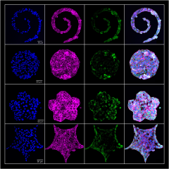
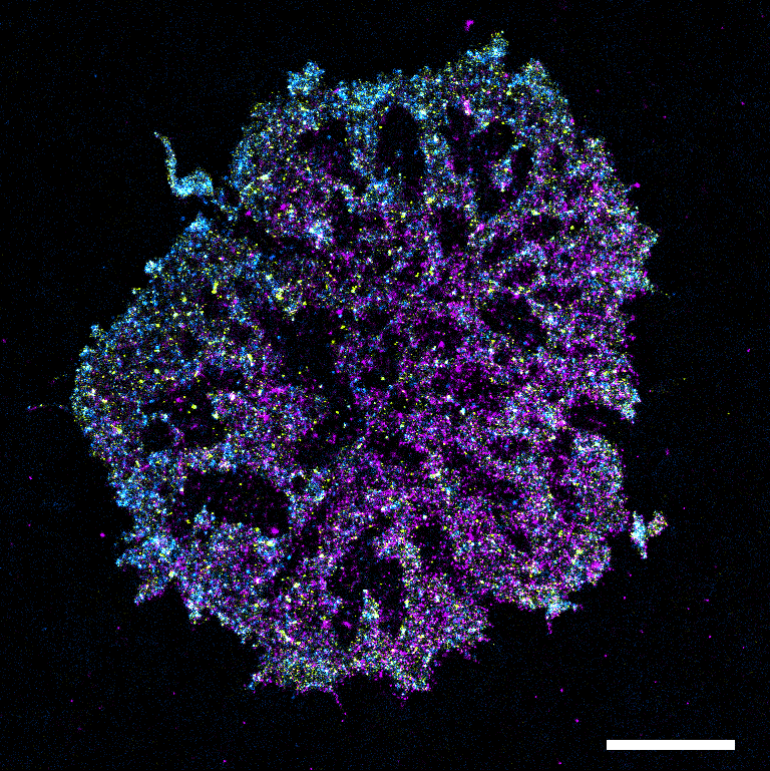
Scroll below to meet the Illuminators and click beneath their profiles to view their art.

Award recipients

Congratulations to the recipients of the winning submissions voted through during UNSW Diversity Festival 2023!

Microscope: Olympus DSX1000 Digital

This video showcases the movement of liquid crystals under an applied voltage signal. Much like the technology used in displays, these unique materials are used and aligned in a special device to detect signals from the heart, brain, and nervous system by converting the electrical signals of these organs into light. It gives us a glimpse into the hidden rhythms that make us who we are, presented in a way that's as enchanting as it is illuminating. 
An Immune Cell’s Response to Intruders

Microscope: Phoebe the Feedback TIRF

In our immune system, the T-cell is always on the lookout for intruders such as infectious pathogens and cancer cells. Upon finding an intruder, proteins on the surface of the cell membrane reorganise to activate signalling processes that ultimately destroy the intruder. 
This is a super-resolution image of an activated T-cell after it recognised an intruder. Each colour represents a different protein that is part of the signalling process.
Can Cells Store Fat?

Microscope: Zeiss LSM 900

Yes, they can! Cells store fats in small pockets called lipid droplets (outlined in green). Lipid droplets are actually made from another compartment within the cells that is essentially a giant network of membrane tubes – the endoplasmic reticulum (purple). When a lot of fat accumulates in the endoplasmic reticulum, it donates part of its membrane to wrap and store it, making lipid droplets. These fat stores can then detach from the endoplasmic reticulum and are used to regulate many cellular processes between different parts of the cell as well as responses to stress. 


Microscope: Nikon A1

Take a peek into the 'Mother Machine,' a fascinating device trapping yeast cells in a family hierarchy. Nestled at the bottom are the 'mothers,' giving rise to a lineage of 'daughters' above them. But what sets this scene alive? It's the glow of green, the spark of energy-producing mitochondria within the cells, passed from mother to daughter with each generation. Fluctuations in mitochondrial partitioning shapes the future of individual cells by influencing growth and successive division. Every split is a fresh start in the 'Mother Machine' where the daughter steps up to become the new mother, continuing the endless cycle of life. 

Gold Nanoparticles Dancing in Green Cell Spheroids

X-ray fluorescence microscopy

We use various techniques to explore how gold nanoparticles enter and spread throughout tight 3D cell clusters. One of these techniques utilises the super-precise X-ray Fluorescence Microscope which can spot and measure metals in biological samples with great accuracy. This allows us to precisely measure the location and number of gold nanoparticles present in cell clusters. Providing valuable insights crucial for the development of nanoparticle-based delivery systems, this research is poised to advance the field of nanomedicine by enhancing the tissue penetration of nanosystems, ultimately leading to more effective disease treatment and diagnosis in the future. 
This image has been adapted from the following publication, with permission: Chen W et al., 'Size-Dependent Penetration of Nanoparticles in Tumor Spheroids: A Multidimensional and Quantitative Study of Transcellular and Paracellular Pathways' (2023), Smallhttps://doi.org/10.1002/smll.202304693.
Exploring Mechanotransduction Pathways in Breast Cancer

Microscope: Leica Stellaris 8

Within the tumour environment, tumour cells encounter an array of forces. Cells make sense of these changes using molecular force sensors which convert these forces to biological signals. These signals allow cells to respond meaningfully to changes in their environment, such as by changing their behaviour. We are particularly interested in one class of force-sensors, the mechanically-activated ion channels, and how they may be involved in regulating interactions between cells. This is relevant in diseases such as cancer where cells can break away from a tumour site and travel through the body to form distant tumours in other organs. 
This image series depicts dissociation of breast cancer cells away from 3D cell clusters over time (from 0- 3 days, left to right). In the top row are normal cell clusters, and in the second row are cell clusters lacking one type of mechanically-activated ion channel. From these images, we can see that deletion of this protein causes more cells to move away from the cluster over time, suggesting a role in regulating cell-cell interactions in breast cancer. In the third row, normal cells and cells lacking a mechanically-activated ion channel have been mixed together so we can see how these different cell types interact.
Uniform Polystyrene Particles

Microscope: Scanning electron microscopy

This image shows how polystyrene forms in the presence of graphene oxide and sodium dodecyl sulfate, through a process called polymerisation where multiple styrene molecules combine together. 
Sucking in the Twenty First Century

Microscope: INCITe Leica Stellaris Dive 

This is what a tongue looks like in a living mouse, where we can visualise the taste buds.
Look Me in the Eye

Microscope: INCITe Leica Stellaris Dive

A microscopic image of the eye cornea from a living mouse! 
Cell Architectural Fireworks

Microscope: Leica Stellaris 8 

We can culture all types of cells from the mouse mammary gland (breast) tissue! Each cell type has a particular shape that is defined by its dynamic skeletal structure – epithelial cells (blue) give functional properties to the mammary gland and are typically rounded, while connective tissue cells such as fibroblasts (magenta + yellow) that support tissue function are typically elongated.
Mammary Tree Sapling

Microscope: Leica Stellaris 8 

When you look under a microscope, the functional cells in the adult mammary gland (breast) form a structure that looks like a tree. When we extract tiny mammary glands from mouse embryos and culture them, we can watch how a simple little mammary tree seed grows into a sapling over a period of 6 days (shown here). This allows me to study the biological processes behind embryonic mammary gland development. 
Fly Talks

Microscope: Leica SP8 

Remember the tiny brown flies hovering over a bygone banana? Here is an image of branches from nerve cells making connections with the chest muscles in our beloved fruit fly. This is how a fly flies!  
The Pit of Despair

Microscope: Leica SP8 

Grooving back at you is the cellular architecture of a Zebrafish eye!
Startling Supramolecular Nanoworms

Microscope: JEOL 1400 

Astonishingly tiny particles (nanoparticles) called worms are developed by the connection between hydrogen molecules. These materials change their shape through heating as the hydrogen connections are disrupted by changes in temperature. Therefore, these temperature-responsive nanomaterials could be used in emerging applications under variable temperature conditions.  

Synthesis of Highly Porous Polymer Nanocomposite Foams Reinforced with Graphene Oxide

Microscope: SEM 230 

Imagine a foam so remarkable that it is not just super lightweight – it is a game-changer.  
Say hello to our cutting-edge foam, meticulously created through a sustainable waterborne technique. This is not your ordinary sponge; it is a marvel of science, boasting high porosity and attractive mechanical properties. 
Smiley Face

Microscope: JEOL1400 

Synthetic substances called polymers can self-assemble into small particles with various forms. We design different kinds of polymers to control the particle shape. This sample happened to turn out to look like a smiley face :) 
Being Conneggted

Microscope: Zeiss 900 

In the ovary, the eggs are fed by the surrounding nurse cells through thread-like processes, called the TransZonal Projections (TZPs). These TZPs feed the egg ions and metabolite goodies. This helps the egg to store the goodies to mature and get ready for fertilization with the sperm. Though, once removed from the ovary, the TZPs don’t last long enough for the egg to mature.  
During in vitro fertilization (IVF), extensive hormonal treatment pushes eggs to mature in the ovary. However, women with certain ovarian disorders and cancer who wish to become mothers are unable to undergo the hormonal treatments.  
 
Our lab has developed a new protocol to help immature eggs to mature in a dish which reduces or eliminates the hormonal treatment needed for IVF. However, the eggs need their goodies. Our research focuses on ways to maintain the TZP numbers to assure the egg receives all the goodies it needs to thrive in a dish! 
Dippin' Dots with Worm Jellies on the Side

Microscope: FEI Nova NanoSEM 450 

This scanning electron microscopy (SEM) image shows different shapes including spheres, worms (fibres), and vesicles formed by self-assembly of special polymers created by Dr. Fumi Ishizuka. 

The Shape of Cancer

Microscope: Zeiss LSM800 

We are interested in how biomechanical cues can affect and induce certain cancer cell subpopulations that drive either tumour progression or trigger metastatic spread of tumour cells to other tissues in the body. Growing melanoma cells on different patterns in the lab allows us to mimic mechanical stress that cells experience in tissues to study how geometric confinement and curvature orchestrate cell response, behaviour and functions. This is exemplified by the green fluorescence signal that is shown to be more prominent on the perimeter of concave patterns.
Atacamite

Microscope: Leica Stereomicroscope

Tabular atacamite crystal surrounded by needles of a translucent, sea-green mineral, likely from the olivenite group. It lays on top of colourless quartz.

Microscope: LSM 900 with Airyscan 2 

Tubulin molecules form the structural foundation of cells. This image depicts tubulin molecules in a human egg. 
In human eggs, tubulin molecules interact with each other to form structural components of the cell and have diverse functions. Tubulins form a network within the cell, creating a superhighway for other molecules and cell organelles to move through the egg. This is essential for egg growth, maturation, fertilization, and overall egg quality.  
Tubulins are also critical to cell division. During cell division, tubulin molecules form rope-like structures and attach to DNA, pulling equal amounts of DNA into the newly formed cells. In our laboratory, we work on ways to improve egg quality to increase a person’s chance of having a healthy baby. 

Microscope: Operetta CLS High Content Analysis System

When treated with chemotherapy, cancer cells can gain compartments called stress granules (green). Stress granules help cells to lower their energy requirements and survive chemotherapy. I model this process in the lab, aiming to understand how stress granules interact with cellular compartments called P-bodies (magenta) and how their interaction may contribute to chemotherapeutic resistance. In my experiments, the cells shrink when exposed to the stress-inducing chemicals. 

Microscope: Zeiss Lightsheet Z1 

How many types of neurons are required for hearing and balance sensory encoding? The diversity of neurons in the inner ear is explored in this study through 3D immunofluorescence microscopy. The two different colours in this image represent two different calcium-binding proteins (green and purple) and thus allow us to visualise two different neuronal types in the spiral and vestibular ganglion. 
Hold Me Close

Microscope: Leica Stellaris 

In the testis, nurse cells called Sertoli cells are critical to the development of sperm. Sertoli cells hold on closely to multiple germ cells at a time and provide them with important nutrients so that they can develop into sperm. Without this important cell type, there would be no sperm. Here you can observe the skeleton of a Sertoli cell in orange still holding on to a few sperm heads in blue.
Supernova

Microscope: Leica SP8 

Multiple melanoma cells cultured together in a ball-shaped clump. The DNA-containing nucleus of each cell is coloured magenta and a protein that forms bonds between the cells is coloured blue.  

Neon Cortex

LaVision Ultramicroscope

This mouse brain underwent a tissue clearing method called CUBIC, where chemicals are used to make the brain tissue transparent (while leaving brain architecture unaltered), resulting in clearer imaging data. This adult mouse was bred to genetically express yellow fluorescent protein in a specific cell type in the brain called pyramidal neurons. The image is focused on the Primary Somatosensory Cortex – the region of the neocortex implicated in the integration and processing of sensory and motor signals.

LaVision Ultramicroscope

This mouse brain underwent a tissue clearing method called CUBIC, where chemicals are used to make the brain tissue transparent (while leaving brain architecture unaltered), resulting in clearer imaging data. This adult mouse was bred to genetically express yellow fluorescent protein in a specific cell type in the brain called pyramidal neurons. The video is a trick of light – it is made up of single images captured through the cortex that are stacked together (a z-stack). When played forward as a video, the light from the microscope illuminates the cells at different levels making it appear as if the cells are firing. This image is focused on the Primary Somatosensory Cortex – the region of the neocortex implicated in the integration and processing of sensory and motor signals. 
Hippocampal Eyebrows on Point

LaVision Ultramicroscope

This mouse brain underwent a tissue clearing method called CUBIC, where chemicals are used to make the brain tissue transparent (while leaving brain architecture unaltered), resulting in clearer imaging data. This adult mouse was bred to genetically express yellow fluorescent protein in a specific cell type within the brain called pyramidal neurons. This image of the hippocampus, the region of the brain involved in learning and memory, is a projection of approximately 80 single images taken through the brain, and compressed together, revealing in 3D the structure and orientation of fibres in this region.  
Motor Neurons in vitro

Microscope: Beta Confocal Microscope 

Image of motor neurons, which are the cells in our nervous system responsible for carrying signals from our brain to our body. Projections responsible for transmitting signals from the cell body to other neurons called axons can be seen branching away from the cluster of cells in the centre. A green and red stain has been used to specifically visualise motor neurons while the DNA-containing nuclei of the cells have been stained with blue. 

Microscope: JEOL JSM-IT500 

Are you tired of coming up with jewellery designs? Take a closer look into nature's realm. Platinum, besides being a noble metal, can create beautiful shapes when it reacts with Gallium. You can see things like flowers and dancing birds. Immerse yourself in the captivating synthesis of science and aesthetics.